Colour vision
Well, we have an understanding of what makes colour, but what makes colour so different from person to person? The answer is in our anatomy - specifically, the anatomy of our eyes. For those of you who haven't done a bunch of anatomy and physiology courses, here's a quick run- down:Light enters your eye through the pupil, which can contract or expand to let more or less of it in (thus making things brighter or darker). That light then gets focused via the lens, which projects the image (upside-down) on the back of the eye, called the retina. The retina is covered in two types of cells - rods and cones.
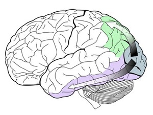
These cells are filled with photosensitive compounds - when enough light causes the compound to break apart, the cell "activates" and sends an impulse down the optic nerve. The occipital lobe of our brain (in the back) along with help from small portions of the temporal and parietal lobes (the specific sections are called the dorsal and ventral streams) translates these impulses into an image.
Most of the "central" image (what you perceive as your focal point) is focused on a small spot called the macula, which is a bit to the outside of the retina. That's because we have stereo vision - the centre of our viewpoint is actually to the inside of each eye's viewing range. Otherwise, we'd have a big problem - the optic nerve, which is in the centre of the back of the eye, has no rod or cone cells on it - it's a small "dead pixel" in our vision.
If you're unsure of what I'm talking about, take a piece of paper and draw a dot on it. Cover one eye, and hold the piece of paper up. When you move the dot so that it is in the centre of your field of vision for the uncovered eye, you'll see that the dot disappears. In order to make up for the "hole" in the vision, your brain will try to fill in the image with the surrounding white paper. Normally, this is avoided by using stereo vision and allowing the central image to focus on the macula of each eye, which is richest in rod and cone cells.


Left: The basic anatomy of the human eye. Rod and cone cells are scattered at the back, where the image is formed;
Right: The occipital lobe, along with help from the ventral (green) and dorsal (purple) streams, help process optical information.
Rod cells respond to the general amount of ambient light, and are responsible for our low-light vision and the overall brightness of what we see. In contrast, cone cells are receptive to one of three main wavelengths of light - red, green or blue (where have we heard that before?). As these cells trip, the brain assigns another level of the corresponding colour to that area of the image, at the brightness dictated by the rod cells.
As you can see, these photosensitive cells are as binary as a computer - they trip or they don't, depending on the quantity of the light they're sensitive to. There is no "in-between," no true analogue quotient. It is just as digital as the display...
...but this is the point where it all starts to go awry.
Colour vision anomalies
Most of us are familiar with the term "colour blind," but have you ever really thought about what it means? Well, what it doesn't mean is not being able to see colour at all, and being stuck with black-and-white vision. Instead, it means an absence of being able to detect one or more (usually just one) of the three basic colours.Human vision is, by and large, trichromatic - we have three cone cells, sensitive to red, green or blue. In a perfect eye, these cone cells are distributed in equal proportion, evenly across the retina. This would allow for even, accurate processing of all colour and intensity of light. Of course, that isn't what happens at all. We already know it's not an even distribution due to the macula having the greatest concentration, meaning that our peripheral vision is inherently less coloured than our central vision.
But let's take that a step further - what if, by some genetic issue, one of the three cone cells didn't form right? You could see red and yellow, but not much blue. Or what if, instead of being sensitive to blue, those cells ended up being sensitive to cyan, instead?


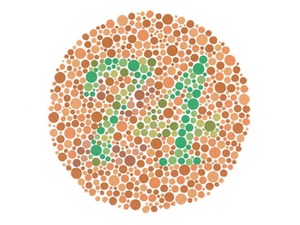
Left: The Ishihara colour test is used to diagnose many colour blindness variations;
Right: The main colours of the rainbow as seen by someone with protonopia, a dichromacy form where red cones are absent.
Colour blindness comes in a variety of forms - there's monochromacy, where at least two of the three cone types flat-out don't work or are not present. Then there's the more common dichromacy, where only one of the three cone types is either absent or malformed enough to not function. Finally, there is anomalous trichromacy, where one of the three receptors is responsive to a wrong wavelength of light - like the cyan example above.
How common do you think that these problems are? Here's a tip - statistically, almost one out of ten of you guys reading this know exactly what one of these is like. Monochromacy is fairly rare, affecting less than one percent of the population. However, the three forms of dichromacy (depending which cone is deficient) affect more than two percent of males. Anomalous trichromacy affects another more than six percent.
Women, on the other hand, are very rarely colour blind. Instead, there's a separate issue that some women have where they are extremely colour receptive, known as tetrachromy. Tetrachromatic women have an entire fourth type of cone cell which is able to pick up yellow light, though those are considerably less prevalent on the retinal surface.

MSI MPG Velox 100R Chassis Review
October 14 2021 | 15:04


Want to comment? Please log in.